

Not All Tubular Adenomas Are from the Intestine
Tubular Adenoma of the Breast
When pathologists hear the term tubular adenoma, the gastrointestinal tract often comes to mind first. However, not all tubular adenomas originate in the intestine. One important — and much less discussed — entity is tubular adenoma of the breast, a rare benign epithelial tumor that can easily be misinterpreted if its features are not well recognized.
In this article, we review the clinical presentation, histopathologic features, differential diagnosis, and clinical significance of tubular adenoma of the breast, with a practical, pathology-oriented approach.
What Is Tubular Adenoma of the Breast?
Tubular adenoma of the breast is a benign epithelial neoplasm composed of tightly packed, uniform tubules. It is considered part of the spectrum of benign breast adenomas and is unrelated to gastrointestinal tubular adenomas, despite sharing the same name.
This lesion is uncommon and accounts for a very small percentage of benign breast tumors.
Clinical Presentation
Tubular adenoma of the breast typically presents as:
- A well-circumscribed, mobile breast mass
- Most commonly diagnosed in young women, often under 40 years of age
- Usually painless and slow-growing
- Often detected incidentally on imaging or during routine examination
Radiologically, it may resemble other benign breast lesions, particularly fibroadenoma, which is why histologic evaluation is essential.
Gross Pathology
On gross examination, tubular adenomas are usually:
- Well-defined and encapsulated or sharply circumscribed
- Firm to rubbery in consistency
- Tan-white on cut surface
- Typically small, though size may vary
Histopathologic Features
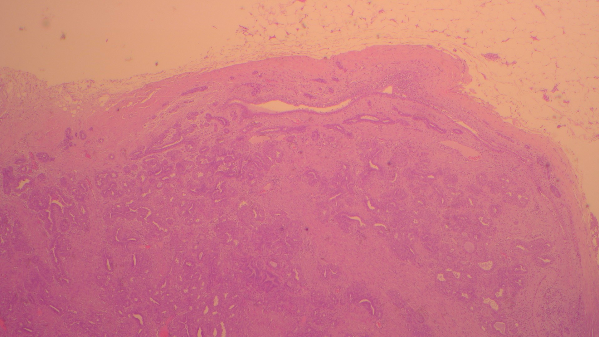
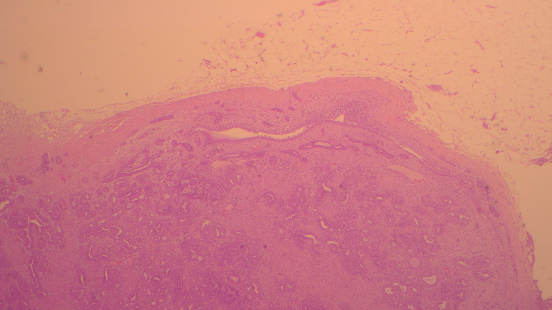
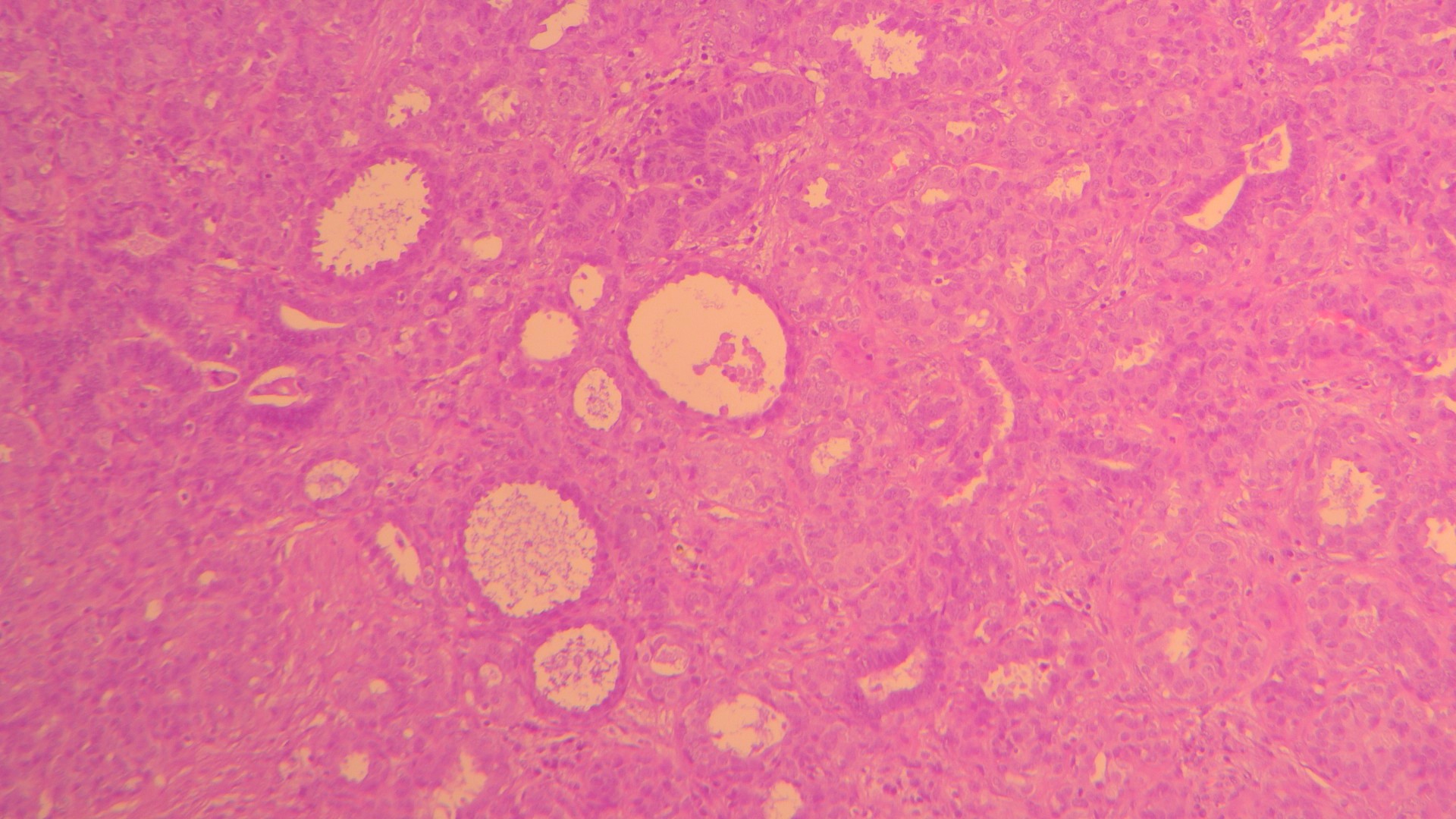
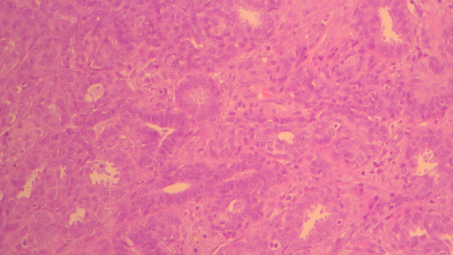
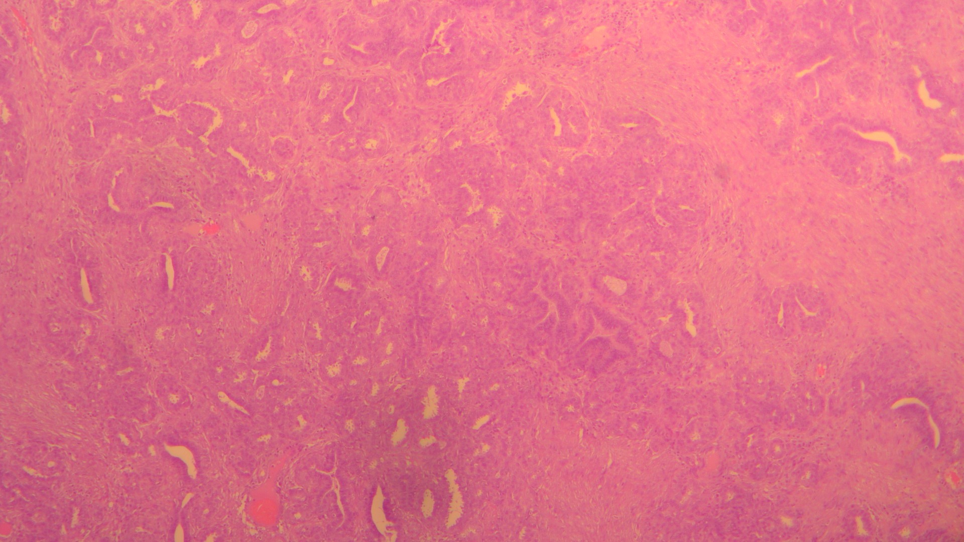
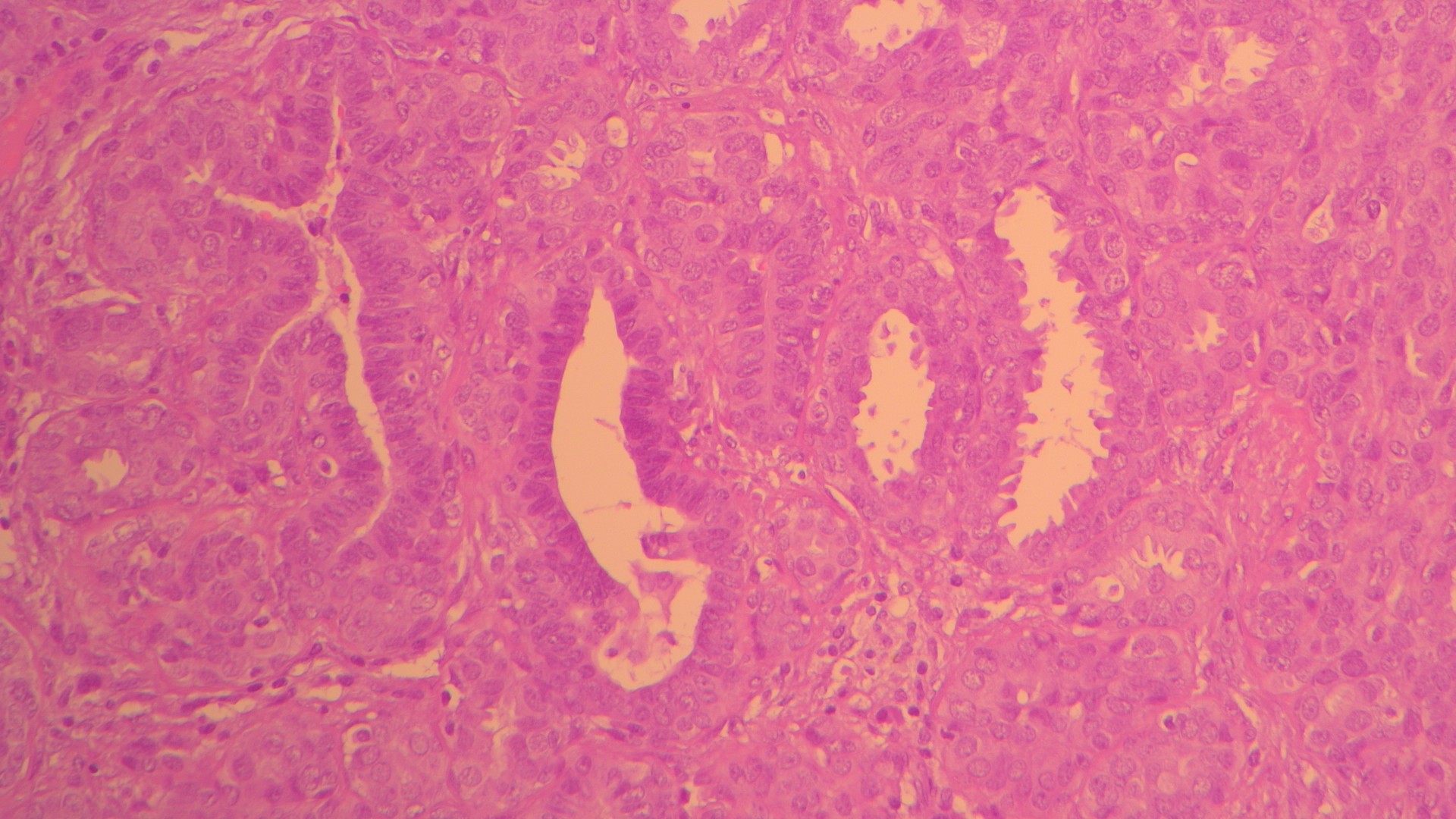
Microscopically, tubular adenoma of the breast is characterized by:
- Closely packed, round to oval tubules
- Tubules lined by two cell layers:
- Inner epithelial cells
- Outer myoepithelial cell layer
- Minimal intervening stroma
- Uniform nuclei with no significant atypia
- Low mitotic activity
The preservation of the myoepithelial layer is a key feature supporting its benign nature.
Immunohistochemistry
Immunohistochemistry can be helpful, especially in small biopsies:
- Myoepithelial markers (e.g., p63, smooth muscle myosin, calponin) highlight the continuous myoepithelial layer
- Luminal epithelial cells typically express cytokeratins consistent with benign breast epithelium
- Low proliferation index (Ki-67)
These findings help exclude invasive carcinoma.
Differential Diagnosis
The main differential diagnoses include:
Fibroadenoma
- More abundant stroma
- Less tightly packed tubules
Lactating Adenoma
- Occurs during pregnancy or lactation
- Shows secretory changes within tubules
Tubular Carcinoma
- Lacks a myoepithelial layer
- Shows infiltrative growth
- Typically associated with desmoplastic stroma
Recognizing the circumscribed architecture and intact myoepithelium is critical to avoid overdiagnosis.
Clinical Behavior and Management
Tubular adenoma of the breast is entirely benign:
- No malignant potential
- Surgical excision is usually curative
- Recurrence is rare
- No association with increased breast cancer risk
Correct diagnosis reassures both clinicians and patients and prevents unnecessary aggressive treatment.
Why This Entity Matters in Pathology Practice
The term tubular adenoma can be misleading. While common in gastrointestinal pathology, its breast counterpart is rare and unfamiliar to many trainees and even practicing pathologists.
Understanding that not all tubular adenomas are intestinal is essential to avoid diagnostic confusion, prevent misclassification as carcinoma, and ensure appropriate patient management.
Final Thoughts
Tubular adenoma of the breast is a rare but important benign lesion that every pathologist should recognize. Its classic histologic features, combined with immunohistochemistry when needed, allow for confident diagnosis and excellent patient outcomes.
In pathology, context is everything — and sometimes, a tubular adenoma is not from the intestine at all.